“Let’s say you have 100 Skittles. If I told you seven of those Skittles could kill you, would you still want to eat any?”
Discovering a potentially life-altering condition is understandably a very scary experience for many. When science teacher Shana Hains received the news concerning her MRI that confirmed the presence of three meningiomas located near her brain and a seven percent chance of going blind, her first response was to call former students.
“Jennifer Wu, who is a neurosurgeon at Harvard [and a former student], responded to my call within minutes,” Hains said. “She walked me through my MRI and told me a little bit about what to expect.”

Meningioma is a tumor that arises from the meninges, or the layers of tissue that protect the brain and spinal cord. Hains first suspected something was wrong when she experienced problems with her vision, a common symptom of meningioma.
She first consulted her optometrist, who found that her optic nerve was swollen, which is why she was recommended for an MRI. Her MRI revealed the tumors, one located on her left optic nerve, another on her frontal lobe, and a third on her brain stem. The sensitive location of these tumors would require very precise treatment.
Fortunately, the meningioma located on Hains’s frontal lobe could be removed with a somewhat simple procedure. This was not the case with the other two.
“Removing the tumor on my optic nerve would be too much of a risky option,” Hains said.
The location of her tumor being so close to the area where her optic nerves crossed meant that removal could cause blindness in not only one, but potentially both of her eyes. Instead of removal, a small cavity was taken out of her skull in order to make room for the tumor. This relieved the pressure it had been applying to her optic nerve and allowed her vision to return back to normal. Unfortunately, she says this surgery was very invasive and required a long, uncomfortable recovery.
“What I hadn’t anticipated was the concussion side effects,” Hains said.
Since her brain had been displaced in order for the surgeons to complete the procedure, she experienced severe concussion symptoms, including fatigue and fogginess.
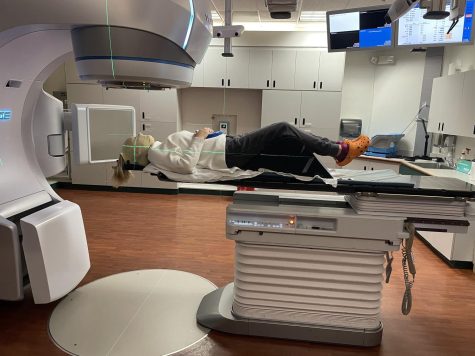
The tumor on her brain stem is located near a cluster of important blood vessels, making removal a very dangerous option. For both the meningioma located on her brain stem and her optic nerve, Hains received 30 days of targeted radiation to kill the cancer cells or stop their growth. This method of treatment uses very narrow intersecting beams of high-intensity radiation to target the tumor more exactly, while also limiting the damage caused to surrounding cells.
“Where the beams cross, the frequency of the waves amplifies,” Hains said.
After 30 days of radiation, Hains said she expects to have regular MRIs to monitor the status of her tumors.
The taxing effects of both surgery and radiation therapy required Hains to take significant time off from teaching. She said that although she felt responsible for her students’ learning, especially her AP Biology class, she knew that the time was crucial for her to recover.
After her return, she spoke positively about the community at West and the support she received.
“I was overwhelmed with the amount of love I received from the staff,” Hains said.
She said that after having taught together for upwards of 20 years, the staff was very tight-knit, and she was grateful for the letters, blankets, cards, and food she received from some of her close friends and coworkers.
Hains mentioned receiving a mixed response from her students, although many were grateful to return to her familiar method of teaching.
“I’ve already started using it as a teaching opportunity in my classes,” Hains said.

